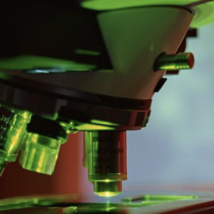
Fluorescent Microscopy: Seeing the Invisible World of Microbes
Fluorescent Microscopy: Seeing the Invisible World of Microbes
If you were to look at a drop of pond water with your naked eye, you’d probably see nothing unusual. But hidden within that drop are countless microorganisms—tiny bacteria, algae, and protozoa—going about their lives. The challenge for scientists is that most of these organisms are invisible without special tools. This is where fluorescent microscopy comes in, allowing researchers to illuminate the unseen and discover the secrets of microbial worlds.
At its core, fluorescent microscopy relies on a simple idea: certain molecules can absorb light at one wavelength and then emit it at another, usually longer, wavelength. This process creates the glow we call fluorescence. By attaching fluorescent tags, often dyes or proteins like the famous green fluorescent protein (GFP), scientists can highlight specific structures within a cell. Under the microscope, what once looked like a blur becomes a dazzling map of color-coded features.
For microbiologists, this technique is transformative. Bacteria, for example, are incredibly small and often look nearly identical under ordinary light. By using fluorescent markers, researchers can distinguish between species, track their movements, or even observe how they interact with one another. In studies of the microbiome—the vast community of bacteria living inside animals, including humans—fluorescent microscopy helps reveal how microbes form colonies, compete, or cooperate.
One fascinating application is in the study of C. elegans, a microscopic roundworm often used as a model organism. Because the worm is transparent, fluorescent microscopy allows scientists to watch bacteria inside its gut in real time. This provides insights into how microbes colonize a host, how they change in response to diet or environment, and how they may influence health.
Beyond biology, fluorescent microscopy has wide-reaching applications. In medicine, it helps detect pathogens, track cancer cells, or monitor how drugs work inside the body. In environmental science, it allows researchers to follow bacteria that break down pollutants. Each glowing image adds to our understanding of life at the smallest scale.
What makes fluorescent microscopy especially exciting for students is its blend of art and science. The images are not only informative but also breathtaking—cells glowing in greens, reds, and blues like miniature galaxies. They remind us that beauty exists even in the tiniest corners of nature, waiting for the right light to reveal it.
In a sense, fluorescent microscopy is more than a tool—it’s a new way of seeing. It transforms the invisible into the visible, the ordinary into the extraordinary. And for young researchers stepping into the lab, it’s a reminder that the world is full of hidden wonders, just waiting to be lit up.
PO-CHENG OU